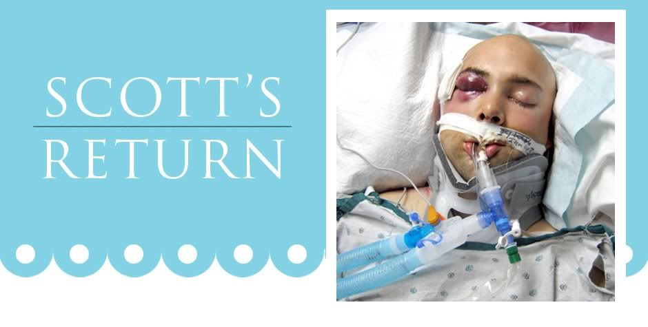
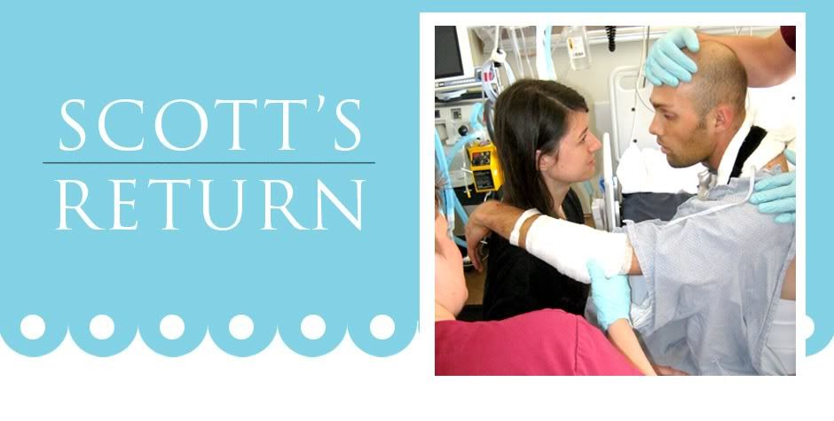
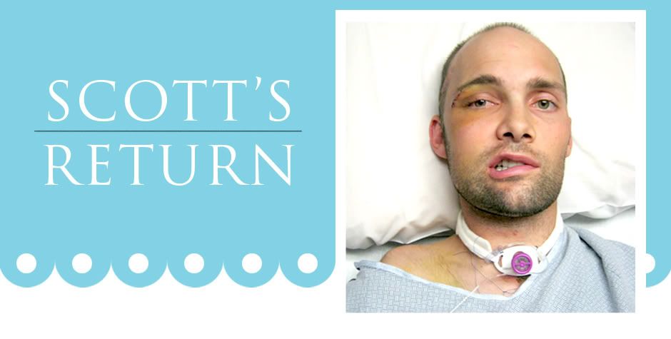
Well,.... so much for a slow day. Guess I should have known better. It is all fine in the end, but ... just some things to deal with.....
Most minor situation first.... As we have mentioned, the external swelling is going down steadily daily. Today, when the nurse was taking care of some wounds on Scott's right shoulder (as you can imagine, major road rash in so many places!), she noticed a big lump on the top of his shoulder. She mentioned it to me, so I went over to look at it. As I was heading over, Ashley mentioned that she thought she saw a clavicle fracture on the x-rays the first day / before I arrived. She did mention it to the doctors, but they were more interested in the life threatening issues in his lungs, appropriately so. Plus, they typically don't do much for a clavicle fracture anyway. After looking at it and knowing the mechanism of injury, I offered that it could be an AC (acromioclavicular) joint separation, and recommended they follow up on it. They pulled up the old x-rays and indeed did find an AC joint separation, so I got to play PT and educate the nurses re: positioning of the shoulder to allow the bones to approximate (be close together), which will optimize healing of the ligaments. Typically, not much more than a sling and physical therapy are done for AC injuries. So... one more owie to get to deal with but nothing major, for sure.
Then, the ICU doc arrived saying that the morning lung x-rays (they take them every day at 4 am) showed some mild infiltrate in the lower left lung pleural space. They wanted to repeat the x-rays with him in a sitting position instead of a supine (lying down) position.
There were 4 things that could possibly be wrong:
1. The current small pneumothorax (in this case caused by a puncture wound in the lung that allows air to escape from the lung into the space between the lung itself and the rib cage. The space shouldn't really be an actual space, but the air creates a space. The membrane around the lungs should actually attach to the rib cage, but if there is air in that space, there is no attachment, so the lungs can collapse. Make sense?) could be growing (like the right one did). Treatment may include a chest tube. One more tube, Yikes!
2. Atelectasis/collapsed lung. Treatment could be done with the ventilator he is already on.
3. Early stages of a mild pneumonia. He has been on antibiotics preventatively since admission, but... still possible.
4. Traumatic Diaphragmatic Rupture, which is caused when the intestines are forced up through the diaphragm during trauma causing a tear in the diaphragm and allowing bowel to penetrate into the pleural space. He did check the original CAT scans, and there was no diaphragmatic rupture noted at that time, but the diaphragm could have been weakened and ruptured later. So.. they wanted to rule it out. I love how attentive they are and don't take anything for granted, even if it feels overwhelming at times to have even one more problem to get to deal with. Surgical treatment would not be an emergency, but may be indicated for him. He would be an "ideal candidate" if it is positive.
They did indeed perform the x-ray and found the following:
1. No traumatic diaphragmatic Rupture. Yeah!
2. Unable to completely rule out the other three, but...not really able to rule them in either. They will keep an eye out, and re-do the x-ray at 3 am.
3. What he thinks is that gravity affected the "air bubble" of the small pneumothorax in the left lung that was already present. So when he was sitting up it was in a different position than when he was lying down. Kind of like the bubble on a level.
Other things of note:
1. Just an observation: he is hyperventilating/taking more breaths than his body needs. Not a concern. It is very common with CNS (Central nervous system) injuries, and may have a protective element to it. Nothing needs to be done about it.
2. The right lung also had a pneumothorax. It was small at first, but did enlarge, so they placed a chest tube in. The right lung has been normal for over 2 days now, so they removed the suction. The tube is still in place, it is just no longer suctioning. If there is a difference between the air pressure in his pleural space and the ambient air, there could still be air movement, but it would not be an active suctioning. So... they removed the suction and will watch it. The lung needs to be normal on it's own for at least 24 hours in order for them to remove the chest tube. That would be awesome to start removing tubes! No more ventilator, no more chest tube... sounds great!
His BP has been running high for the first time....just since the surgery. Keeping an eye on that, too.
Oh... and the MD mentioned today that he wants him still for 24 hours after the surgery to allow the seals on the trach tube and the PEG tube to heal well. So... his sedation will be kept high and they won't start "wake assessments" until tomorrow afternoon.
Have a good night's sleep, and we'll "talk" again in the morning!
We love you!
Patti


No comments:
Post a Comment